 |
| Ameloblastic fibroma showing epithelial and mesenchymal components. |
sábado, 27 de mayo de 2017
Ameloblastic fibroma
Interestingly, new 2017 WHO/IARC classification refers that ameloblastic fibroma may show formation of dental hard tissue, as ameloblastic fibrodentinoma or ameloblastic fibro-odontomas are no longer part of the odontogenic tumor classification.
I reckon (a daring personal opinion) a vast evidence of tumor genetic profiles are needed to separate odontogenic lesions in a new classification or, even worse, eliminate them for good. Opinions?
Odontogenic keratocyst
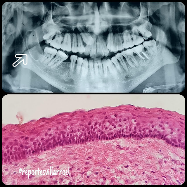
Reclassified again as odontogenic cyst WHO 2017 (former odontogenic keratocystic tumor).
 |
| Fig. 1 . X-ray showing typical anterior-posterior cyst growth. Microscopic pic shows representative features of epithelial lining of keratocyst. |
 |
| Fig. 2. Cyst with a lined by a parakeratinized stratified squamous epithelium with tendency to detach from the connective capsule. |
Suscribirse a:
Comentarios (Atom)
